What is radioembolization?
Radioembolization treats liver cancer by delivering tiny radioactive microspheres directly into the blood vessels feeding a tumour. Guided by a thin catheter inserted through a small incision (usually in the groin or wrist), the beads lodge in the tumour and release a high, localised dose of radiation while protecting the rest of the liver.
What it treats
It is used for primary liver cancer (hepatocellular carcinoma) and for cancers that have spread to the liver from elsewhere — such as colorectal, breast or neuroendocrine tumours — and is especially valuable when surgery is not possible because of the tumour's size, location or the patient's health. It can sometimes shrink a tumour enough to make surgery possible later (a “bridge to surgery”).
What to expect
Treatment usually involves two visits a few weeks apart: a planning angiogram to map the blood supply, then delivery of the microspheres through the catheter under real-time X-ray guidance. The procedure takes one to two hours and is generally not painful thanks to local anaesthetic, with light sedation available for comfort.
Advantages
- Concentrates radiation on the tumour while sparing healthy liver
- Minimally invasive, with a shorter stay and faster recovery
- Fewer whole-body side effects than traditional chemotherapy
- May open the door to curative surgery or transplantation
Good to know
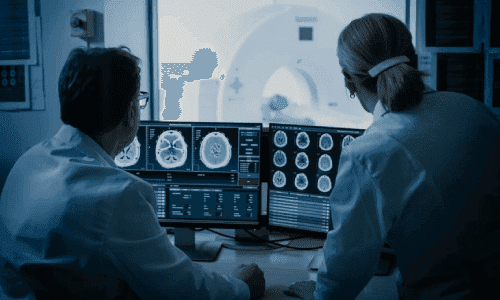
It is not suitable for everyone — for example with severe liver failure, widespread disease or pregnancy. Some people get temporary “post-embolization syndrome” (fatigue, mild fever, nausea, abdominal pain) that settles within a week. You will need someone to drive you home, should limit close contact with children and pregnant women for a few days, and will have follow-up scans to check the response.