What is PET-MR?
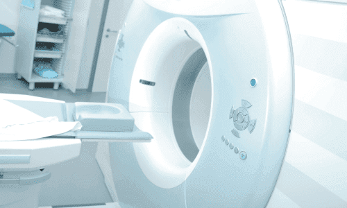
PET-MR merges two technologies. PET shows the body's metabolic activity — how cells are working — and highlights areas that may signal disease. MRI provides clear, detailed images of organs and soft tissue using magnetic fields, not X-rays.
Captured together, they produce a single fused image showing both what an abnormality is and how it is behaving — leading to more accurate diagnoses and better-planned treatment, especially in oncology, neurology and cardiology.
Key features
- Superb soft-tissue detail for precise lesion detection
- Much lower radiation than PET-CT, as MRI uses no X-rays
- Functional and anatomical data captured in one session
What it is used for
In oncology, PET-MR detects and stages tumours and checks how treatment is working. In neurology, it helps assess conditions such as Alzheimer's disease and locate the source of epileptic seizures. In cardiology, it evaluates heart-muscle health and blood flow.
What to expect
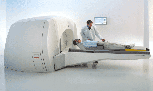
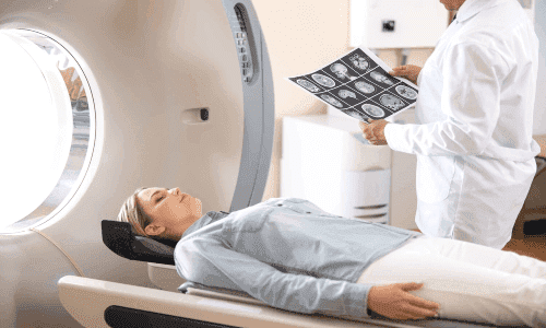
A low dose of a radioactive tracer is injected, usually into a vein — the only sensation is a small needle prick. After a quiet rest of 45–90 minutes while the tracer circulates, you lie still inside the scanner for 30–60 minutes. The whole visit takes about 1–2 hours and is otherwise painless.
Good to know
Because of the strong magnet, PET-MR may not suit people with a pacemaker or certain metal implants, and you should tell your team if you might be pregnant. Serious side effects are extremely rare; drinking plenty of water afterwards helps clear the tracer, and you can return to normal activity straight away.