What is EOS imaging?
EOS uses a biplanar, slot-scanning technique — two narrow X-ray beams scan the body simultaneously to capture front and side views while you stand or sit naturally. Based on Nobel Prize-winning detector technology, it does this with far less radiation than conventional X-rays or CT.
Key features
- Very low radiation — 50–85% less than conventional X-ray
- High-resolution 2D and precise 3D skeletal models
- Imaging in a natural, weight-bearing position
- A full-body scan from head to toe in a single pass
What it is used for
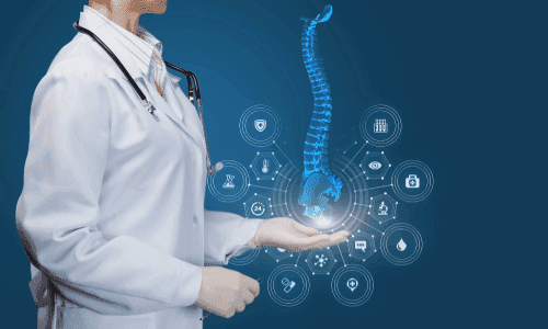
EOS is ideal for assessing and monitoring scoliosis and other spinal deformities, leg-length differences, and hip and knee alignment, as well as planning orthopaedic surgery and checking results afterwards.
What to expect
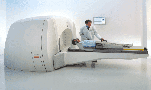
You stand or sit still inside the scanning cabin for just a few seconds while the system captures the images. A full-body scan takes about 20 seconds and is completely painless and non-invasive — no needles, no enclosed tunnel.
Good to know
Its very low dose makes EOS especially valuable for children and anyone needing regular monitoring. It is avoided in pregnancy, and clear images depend on staying still for a few seconds. There are no known serious side effects, and you can return to normal activity straight away.