What is 3D mammography?
3D digital breast tomosynthesis captures multiple low-dose X-ray images from different angles to build a layered, three-dimensional map of the breast. Unlike flat 2D images, where overlapping tissue can hide a problem, it lets radiologists examine the breast in thin, distinct slices — much like reading a book page by page.
Key features
- Layered images that remove the confusion of overlapping tissue
- Fewer call-backs and less unnecessary follow-up testing
- Better detection in dense breast tissue
- Earlier detection of small, early-stage cancers
What it is used for
It is used for breast-cancer screening and early diagnosis — finding small abnormalities, investigating lumps or breast pain, and performing especially well in women with dense breast tissue, where 2D mammography can be less reliable.
What to expect
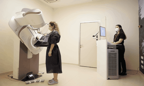
You stand at the unit and the breast is gently compressed for a few seconds for each image — this can feel like brief pressure but is generally not painful. The whole appointment takes about 10–15 minutes. Please avoid deodorant, perfume or lotion on the day, as these can affect the images.
Good to know
It uses a very low dose of radiation, similar to a standard 2D mammogram and well within safety limits — the benefit of early, accurate detection far outweighs this minimal exposure. It is not used in pregnancy. No special aftercare is needed and you can return to normal activity immediately.