
ما هو مرض القلب الخلقي؟
وعندما لا يتطور قلب الطفل عادة في الرحم، تحدث اضطرابات هيكلية ووظيفية. ويتطور قلب الطفل في الأسبوع الثامن من الحمل. هناك أنواع عديدة من أمراض القلب الخلقية، من بسيطة إلى معقدة، ووظيفة الشاذ
It is when the baby’s heart does not develop normally in the womb and structural and functional disorders occur. The baby’s heart is developed in the 8th week of pregnancy. There are many types of congenital heart diseases, from simple to complex, and the function of the abnormally structured heart and its ability to pump blood to the body can be seriously affected. Babies’ health, growth, and development may be affected to varying degrees. Congenital heart disease is seen in approximately 0.8% of every 100 live-born babies in Turkey. The development of technologies used for diagnosis, monitoring of these processes, and advances in surgical interventions allow babies with congenital heart disease to be diagnosed earlier and live a longer and healthier lives. It is recommended that babies with congenital heart disease be kept under the control of a professional team.
Anomalies in the baby’s heart can be detected early with fetal echocardiography performed during pregnancy. If congenital heart disease can be detected intrauterine (in the womb), family members can be informed to gain time to understand the diagnosis, evaluate fetal anomalies, and decide on the continuation of the pregnancy based on personal values. For families who want to continue the pregnancy, it is vital that doctors and the family plan fetal monitoring, deliver safely and on time, and choose a fully equipped hospital where medical care that may be needed immediately after birth can be provided.
ما هي أعراض أمراض القلب الخلقية؟
وتبعاً لنوع وشدة أمراض القلب الخلقية، قد يعاني الأطفال من أعراض مختلفة كثيرة. بعض المرضى قد يكون لديهم أعراض قليلة أو لا In symptomatic patients, bruising on the skin, lips, and nail bases, blisters on the fingers, respiratory distress, recurrent lung infections, difficulty in absorbing, inability to gain weight, excessive wing while spraying, shortness of breath, and gettingted easily while moving or playing may be observed.
ما هي أسباب أمراض القلب الخلقية؟
Congenital heart disease occurs due to developmental disorders that occur in the womb during the first 8 weeks of pregnancy. Although the exact cause is unknown, some maternal factors may predispose to the development of congenital heart disease. Some medications used by the mother play a role in the development of congenital heart disease. For example, isotretinoin and anticonvulsant drugs used in the treatment of acne can be listed. It is known that some viral infections during the first 8 weeks of pregnancy may also play a role in the development of congenital heart disease. However, genetic anomalies may increase the risk of developing congenital heart disease. The most common genetic pathologies are Down syndrome, Turner syndrome, Marfan syndrome, and Di-George syndrome. The probability of developing congenital heart disease in pregnancies using assisted reproductive techniques and in babies of parents with a family history of congenital heart disease increases, albeit slightly. That’s why families need to receive genetic counseling before pregnancy.
كيف يتم تشخيص أمراض القلب الخلقية؟
It can generally be diagnosed by prenatal fetal echocardiography, but in our country it is mostly diagnosed by postnatal transthoracic echocardiography (TTE). Very rarely, the diagnosis can be made at a later age due to the child’s lack of signs and symptoms. Procedures performed for diagnosis include ECG (electrocardiography), echocardiography (ECHO), angiography, magnetic resonance imaging (MRI), computerized tomography (CT). Some heart anomalies may be difficult to detect at an early age and may not be diagnosed until adulthood. The diagnosis of these patients depends on the development of complications. Besides ECG and ECHO, the most commonly used device for diagnosis is pulse oximeter. Thanks to this device, the oxygen saturation in the baby’s fingers and toes is simply measured, and the performance of the heart and the amount of oxygen in the blood circulating in the body are measured.
من هم أعضاء فريق أمراض القلب الخلقية؟
ويوصى بإجراء جراحة مبكرة للأطفال الذين يحتاجون إلى جراحة القلب خلال فترة الولادة الجديدة. وفي المريض المشغّل، ينمو القلب مع الطفل، وتتحقق عملية تعافي أسرع خلال الطفولة. ويجري تنفيذ أكثر من ثلثي عمليات القلب والأوعية الدموية في مرحلة الطفولة على المرضى الذين تقل أعمارهم عن سنة واحدة. آلة القلب تستخدم في معظم هذه العمليات وهذه الطريقة تجعل إجراءاتنا الجراحية أكثر كفاءة وأمنا. ويطبق برنامج علاجي فريد على كل مريض. ولهذا السبب، من الأهمية بمكان إجراء الجراحة اللازمة في الوقت المناسب مع التشخيص والتقنية والفريق الصحيح. ويقوم تشخيص أمراض القلب الخلقية أخصائي أمراض القلب، الذي يشكل جزءا لا يتجزأ من الفريق. وبعد القيام بالتدخلات اللازمة على المريض من قبل أخصائي أمراض الأطفال في غرفة العمليات، يقوم الجراحة جراحة طبيب الأطفال. آلات القلب المستخدمة أثناء الجراحة تدار من قبل شخص ذو خبرة في جراحات القلب وعندما تكتمل الجراحة، يُنقل المريض إلى وحدة الرعاية المكثفة ويُعهد به إلى فريق ذي خبرة يشمل أخصائيي القلب والممرضات والأطباء المتخصصين في الرعاية المكثفة.
معالجة أمراض القلب الخلقية
Interventions that can be performed for the treatment of congenital heart patients include drug therapy, interventional angiography, and heart surgery. Although some congenital heart diseases do not require treatment, intervention is often required in children with clinical complaints. For this reason, we serve all our patients every day, every hour in our hospital. We apply all treatment methods used in cardiac surgery. NIRS (Near-Infrared Spectroscopy) device, which provides close monitoring, is used to monitor and protect our patient’s brain functions during heart surgery. Except for procedures that require a sterile environment, care is taken to ensure close contact between the family and the baby.
إغلاق العيوب التخديرية من خلال شق أدنى
هدفنا هو تشخيص وعلاج مرض القلب في أقرب وقت ممكن. وهدفنا الرئيسي هو معالجة الطفل في أقرب وقت ممكن وإعادة الطفل إلى الحياة الطبيعية قبل أن يبلغ سن المدرسة. While some congenital heart diseases can only be treated with medications and monitored by a pediatric cardiologist, some complicated heart diseases can also be treated with cardiac catheterization (angiography) and sometimes wound at a very early stage. وهكذا، يمكن أن يعيش الأطفال حياة طبيعية بعد الجراحة أو العلاج الأوقياني. غير أنه في أمراض القلب المعقدة الأخرى، يمكن تطبيق بروتوكولات العلاج المشتركة، وقد يلزم إجراء عدة جراحات قلبية مرحلية. لدينا فريق كبير يعالج كل مريض على حدة ويحدد أفضل علاج للطفل بالنسبة لبعض أمراض القلب الخلقية الشديدة قد يحتاج الأطفال إلى جراحة بعد الولادة مباشرة إن العلاج المبكر والمتابعة أمران ضروريان لأن فقدان الوقت قد يتسبب في فوات فرصة الجراحة.
Echocardiography (ECHO)
Echocardiography is an imaging method that obtains a two-dimensional and three-dimensional real-time image of the heart using sound waves. ويتم الحصول على معلومات وقياسات بشأن التشريح ووظيفة القلب. ويتم جمع الأمواج الصوتية العالية التردد التي تكون عالية جداً بحيث يستمع إليها الأذن، التي تُرسل إلى قفص الصدر من خلال الجهاز، عندما تعود من الجسم، ويتم الحصول على قياسات تعطي فكرة عن مظهر القلب ووظائفه. وبعبارة أخرى، فإن صدى القلب هو حاسوب يحول موجات الصوت المرسلة إلى القلب إلى صور. ويمكن القيام بذلك في أي وقت طوال الحياة، بما في ذلك فترة الرحم. ويُستخدم الفحص الأوقيانوغرافية استخداماً فعالاً كأسلوب مساعد للتصوير أثناء التشخيص في تشخيص مشاكل القلب، وفي تخطيط الجراحة قبل جراحة القلب، وفي تقييم عمليات المتابعة في فترة جراحة ما بعد القلب.
Types of echocardiography are Transthoracic Echocardiography (TTE), Transesophageal Echocardiography (TEE), Fetal Echocardiography, 3D Echocardiography, Stress Echocardiography, Contrast Echocardiography, Intravascular Echocardiography. Transesophageal echocardiography is an echocardiography method based on imaging the heart from inside the body through a specialized transducer placed in the esophagus. In TEE, sound waves do not have to pass through the skin, muscle, and bone tissue, resulting in a much clearer image. Sedation is always used during TEE. Sedation may sometimes be required in young children due to poor patient compliance. In this way, patient compliance is increased and image quality and measurements can be made in the most accurate way. There is no pain during echocardiography because echocardiography works with sound waves, but sometimes pressure on the chest cage to increase image quality can cause mild pain and discomfort in children. In addition, the coldness of the gel applied and the tickling when touched may cause disharmony and restlessness in the child. Echocardiography is not harmful to health, it works only with sound waves and does not emit any radiation. For this reason, it can be used safely both during pregnancy to check the baby’s heart and in children.
Angiography
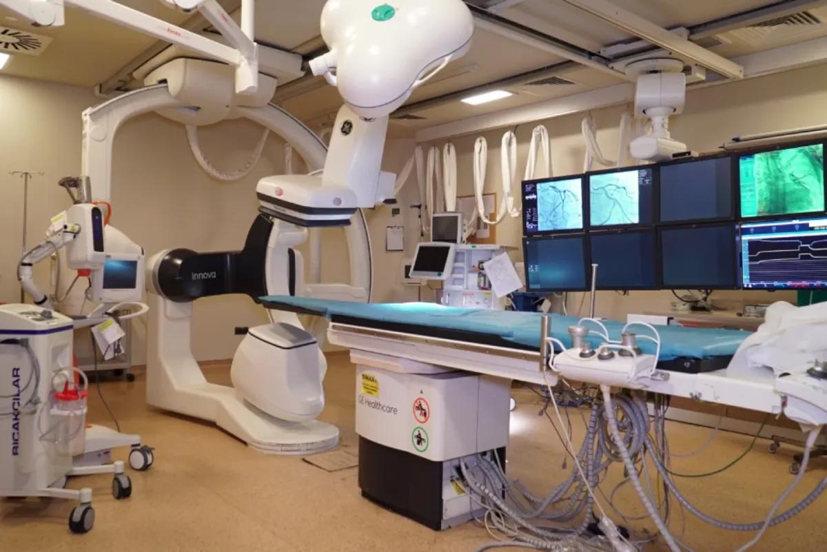
وتتم الوصول إلى غرف القلب والأوردة من خلال التقدم عبر الوريد من خلال الأسلاك الرقيقة والأنابيب ذات السمات التقنية العالية. وهنا، يتم أخذ قياسات الضغط وعينات الغاز الدمي وإجراء الحسابات. ومرة أخرى، يكشف التشريح عن طريق تطبيق مادة متناقضة على هذه المجالات. غير أنه يمكن أيضا تنفيذ إجراءات التدخل لأغراض العلاج. إن إزالة البذور عن طريق تضخم بالون في مناطق ضيقة، وإغلاق الفتحات في جدران القلب بأجهزة غير حصرية خاصة، أو وضع مخزن في السفينة يحتاج إلى البقاء مفتوحاً، يمكن أن يتم عن طريق الأوقيانوغرافية. ويجري تنفيذ الإجراءات المتعلقة بعلم الأطفال في إطار التخدير العام بسبب الحاجة إلى الامتثال الكامل للمرضى. Angiography is the method used to identify or resolve complications and pathologies before or after operate, diagnostic or interventional.
التصوير الصوتي المغناطيسي
ويعتبر التصوير بالرنين المغناطيسي القلبي من أسرع الأدوات التشخيصية نمواً في مجال طب الأطفال. وهي تتيح الفرصة للحصول على صور ثلاثية الأبعاد وصور عالية الاستبانة عن تشريح القلب والمرض المتقدم. وهو أسلوب غير متفشي وخالي من الإشعاع. وكثيراً ما يتم ذلك تحت التخدير بسبب طول فترة إطلاق النار والحاجة إلى امتثال المرضى. وتتمثل مزاياها الرئيسية على أساليب أخرى في أنها توفر صورة حقيقية عالية الاستبانة عن التشريح والعلاقة مع الأجهزة المحيطة بصورة ثلاثية الأبعاد، ويمكن إجراء دراسات التدفق، وأنها غير متفشية ولا تحتوي على الإشعاع.
الترجيح المحوسب
وبالمثل، فإنها تتيح الفرصة للحصول على صور ثلاثية الأبعاد ومرتفعة الاستبانة غير متفشية. وتتمثل أكبر ميزة لها في أن لديها حلاً أعلى ووقتاً أقصر من وقت الاحتياز مقارنة بالرنين المتعدد الأطراف. وحتى إذا كان الترسيب ضروريا، تطبق بروتوكولات قصيرة الأجل. لكن التعرض للإشعاع هو أهم عيوبه وقد انخفض مستوى الإشعاع انخفاضاً كبيراً في أحدث أشعة مقطعية متطورة اليوم.